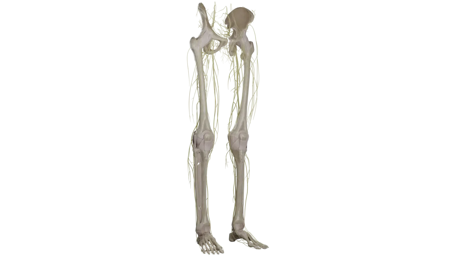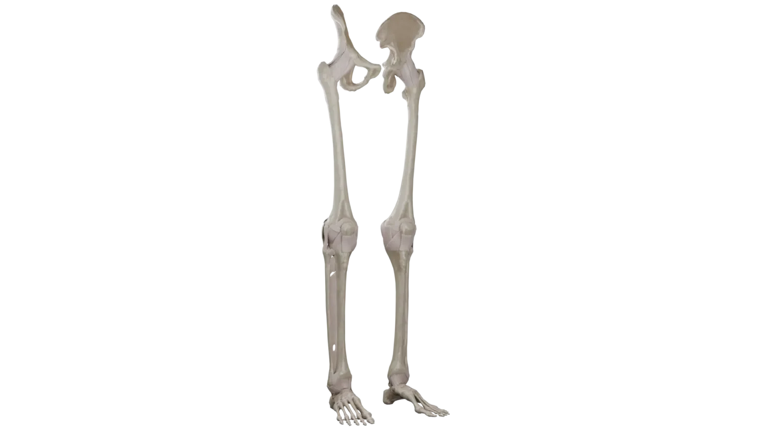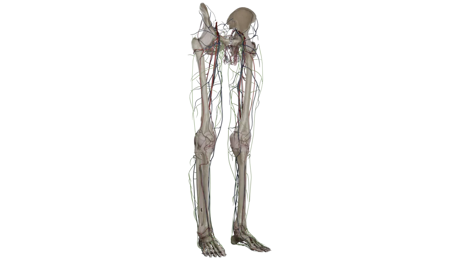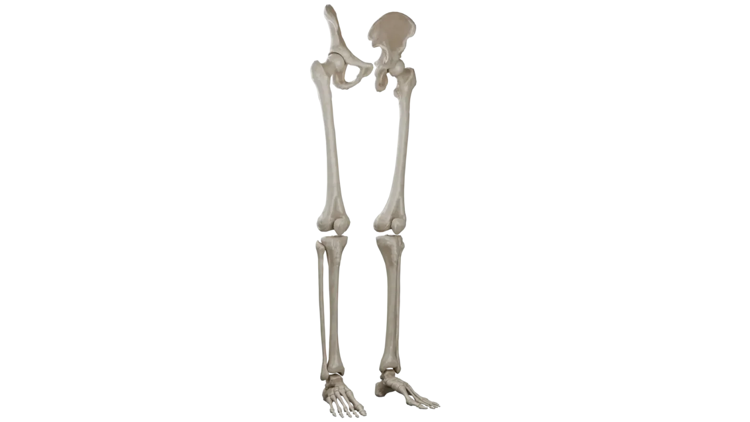
Skeletal anatomy of the lower limb includes the pelvic bones, femur, patella, tibia, fibula, and the bones of the foot. The pelvic girdle, formed by the ilium, ischium, and pubis, supports the weight of the upper body and provides attachment points for muscles. The femur, the longest bone in the body, articulates with the hip and knee joints. The tibia and fibula form the skeleton of the leg. The foot comprises the tarsal bones, metatarsals, and phalanges, enabling complex functions. Detailed 3D models present the shape, articulations, and structural features of these bones, crucial for understanding the biomechanics of the lower limb.
Show more Show less