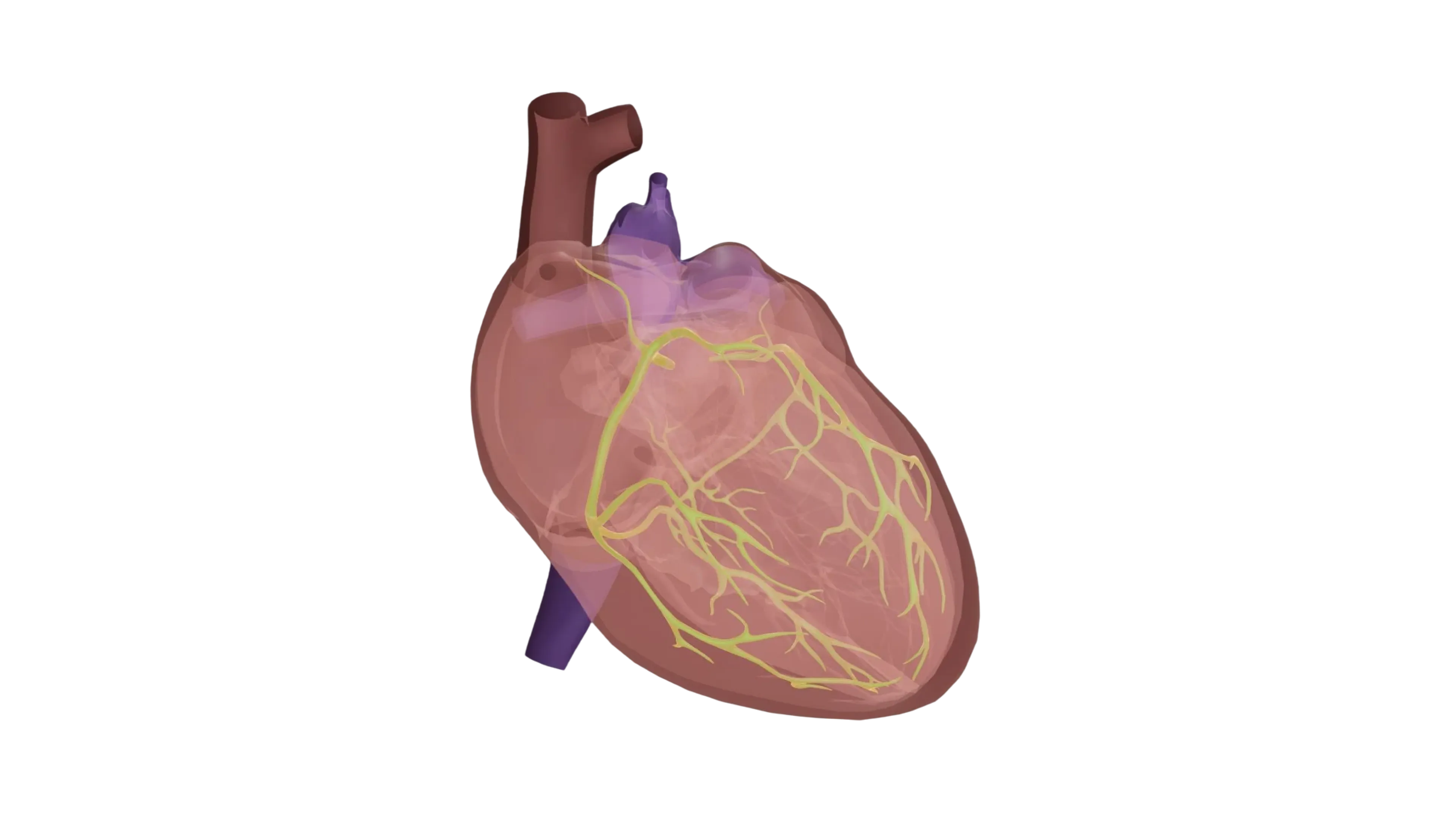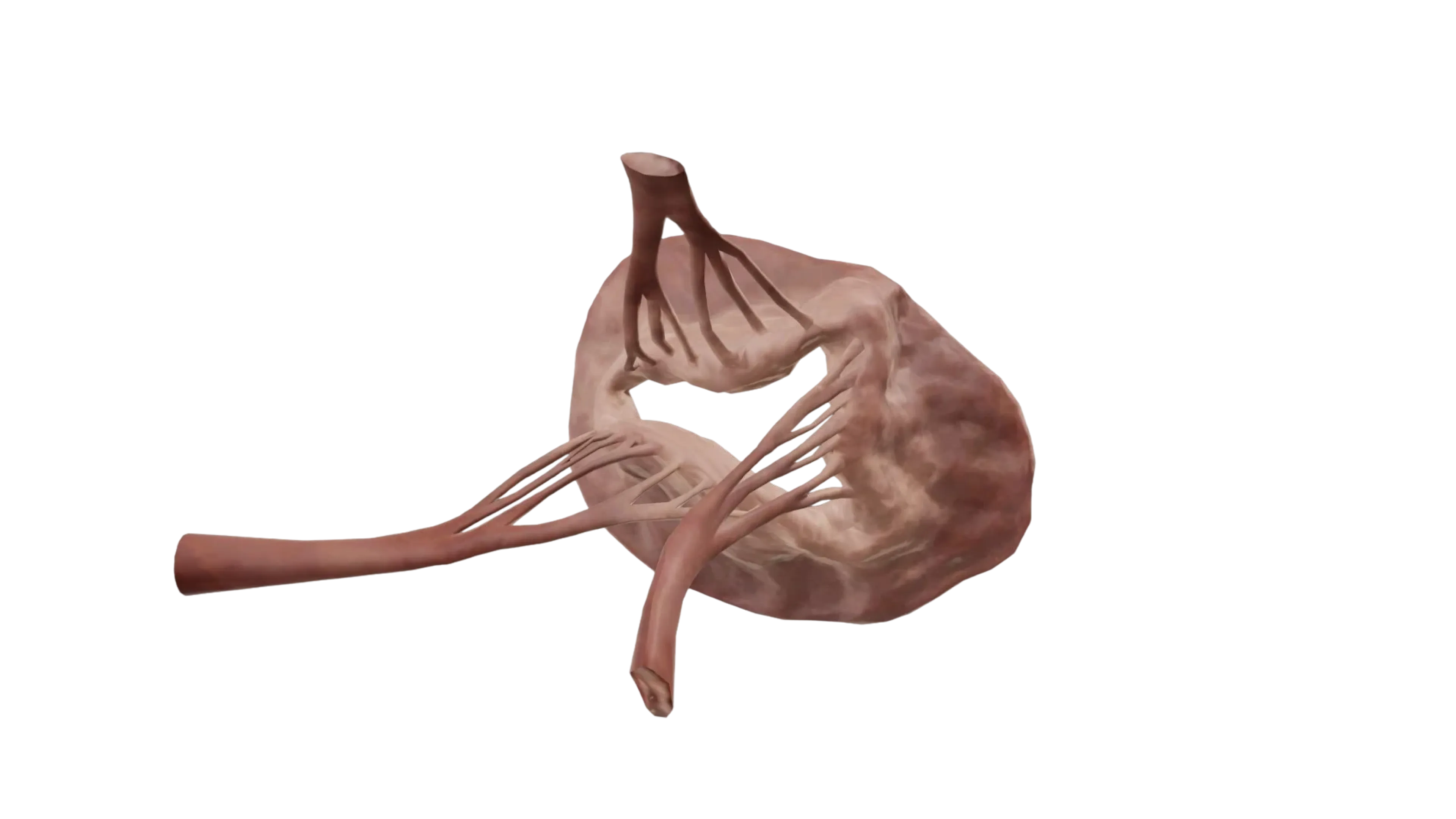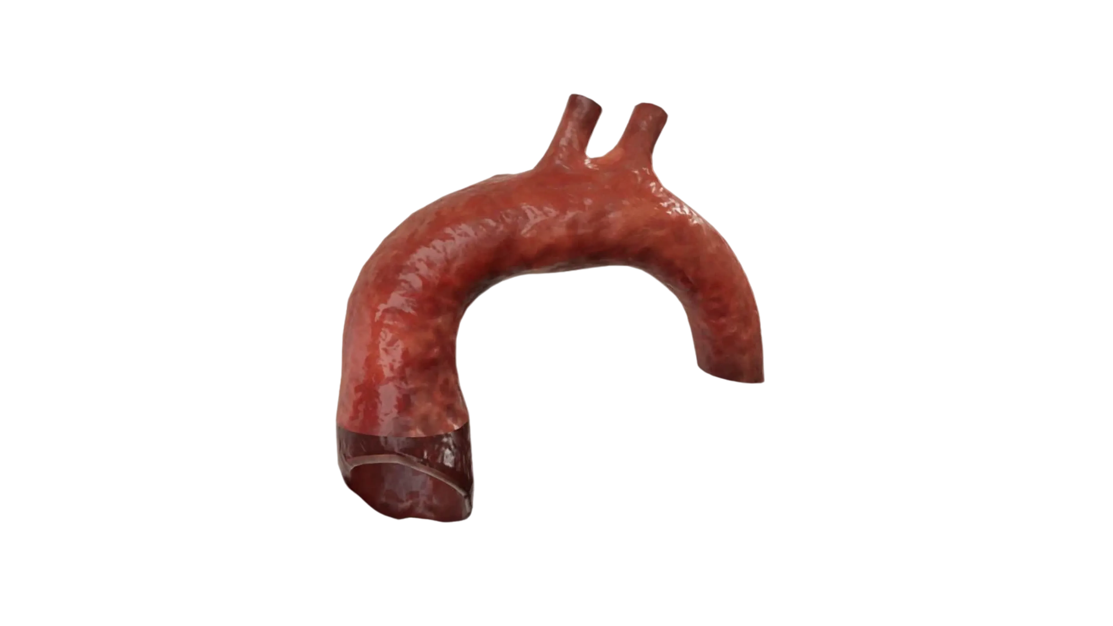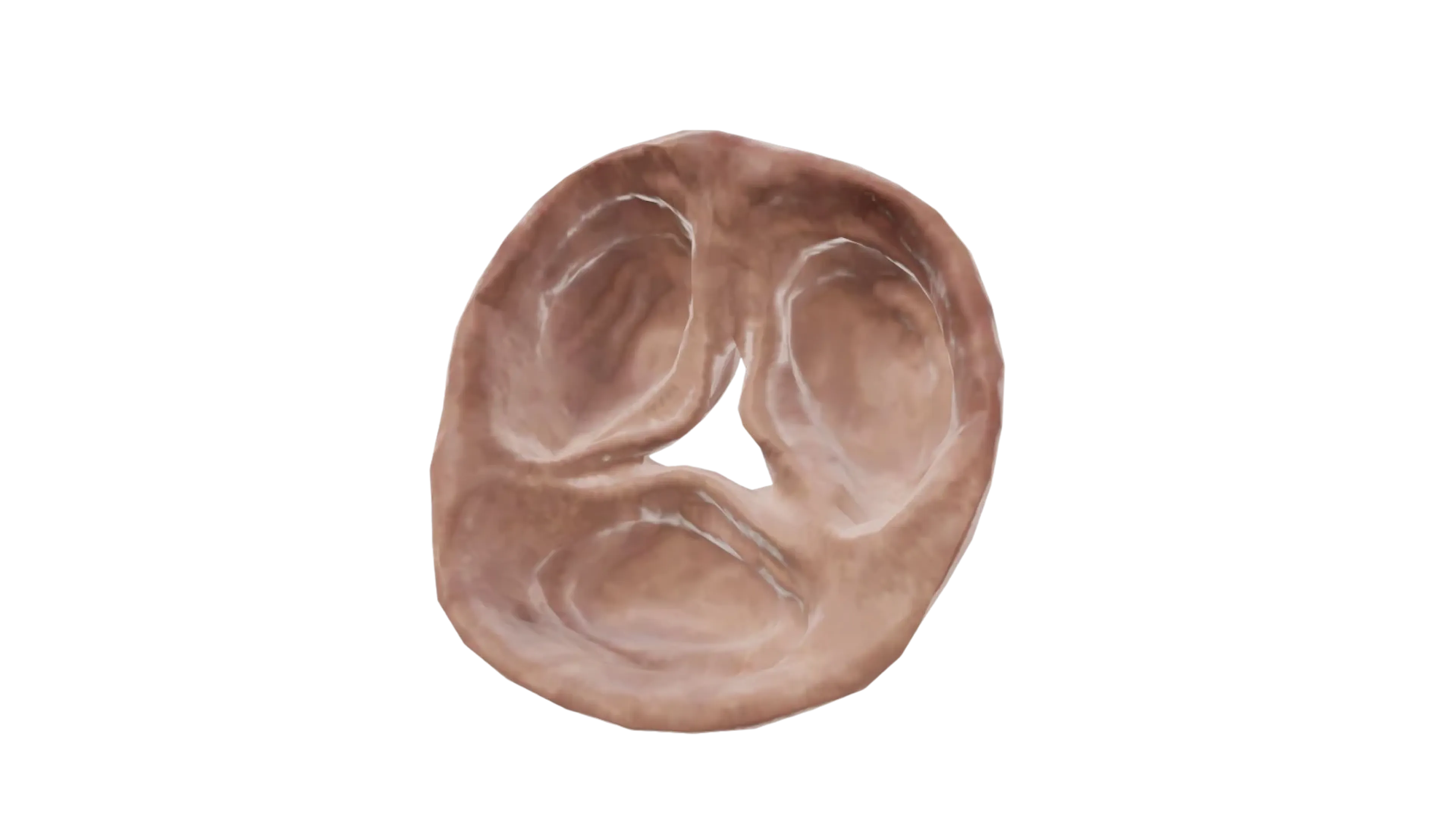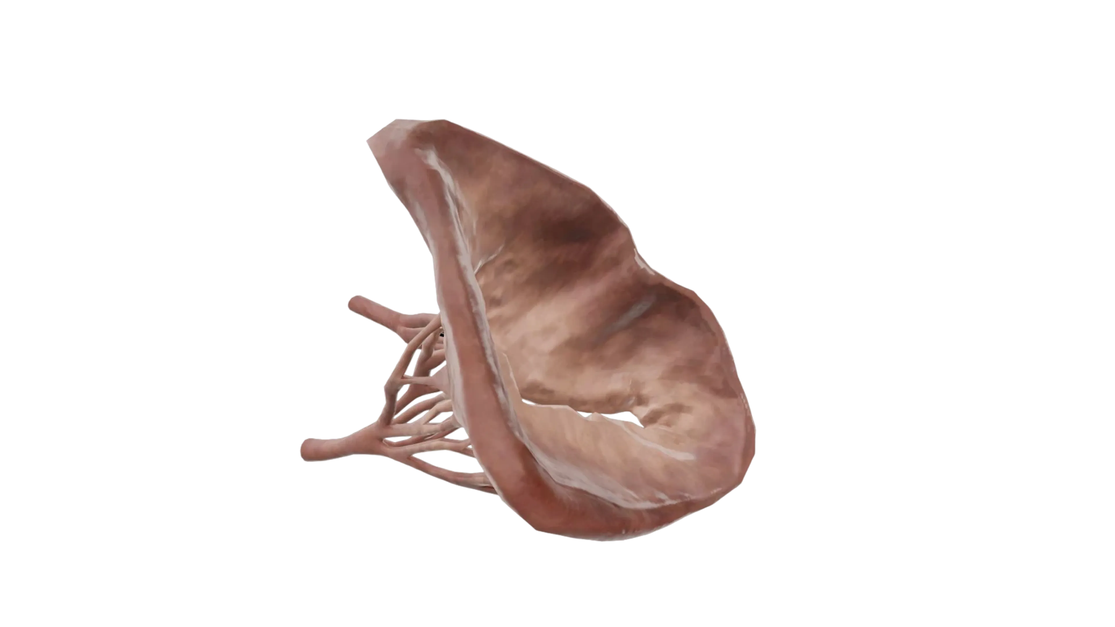
Examine the intricate structure of the mitral valve with this 3D anatomy model. It illustrates the anterior and posterior leaflets, chordae tendineae, and papillary muscles, providing a realistic representation of valve function during the cardiac cycle. Medical students and professionals can use the model to understand normal blood flow from the left atrium to the left ventricle and enhance comprehension of surgical and diagnostic approaches in a three-dimensional, interactive format.
Show more Show less