
3D pathology models: new lens on understanding diseases
Share:
Copy link
Share via Telegram
Share on Facebook
Share on LinkedIn
Share via WhatsApp
Share on X
Share via Email
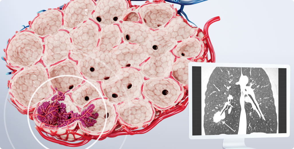
Table of contents
Studying diseases at every level, from cells to entire systems, is crucial for accurate diagnosis and effective medical research. However, traditional pathology visualizations offer only a flat, single-plane view, often missing the full complexity of biological systems. In this article, we explore how 3D pathology models overcome the limitations of 2D imaging and drive advancements in the healthcare sector, including enhancements in pathology and pathophysiology learning, patient education, and clinical preparations.
What is a 3D pathology model?
3D pathology models are advanced medical representations that allow researchers and practitioners to study diseases and disorders in three dimensions. This approach offers a more comprehensive and realistic view compared to traditional 2D imaging.
Such models capture the complexity of biological systems, including the spatial relationships between cells, tissues, and structures. The wider perspective offers deeper insights into disease mechanisms and potential treatments.
There are different types of pathology models, with each one serving specific use cases:
-
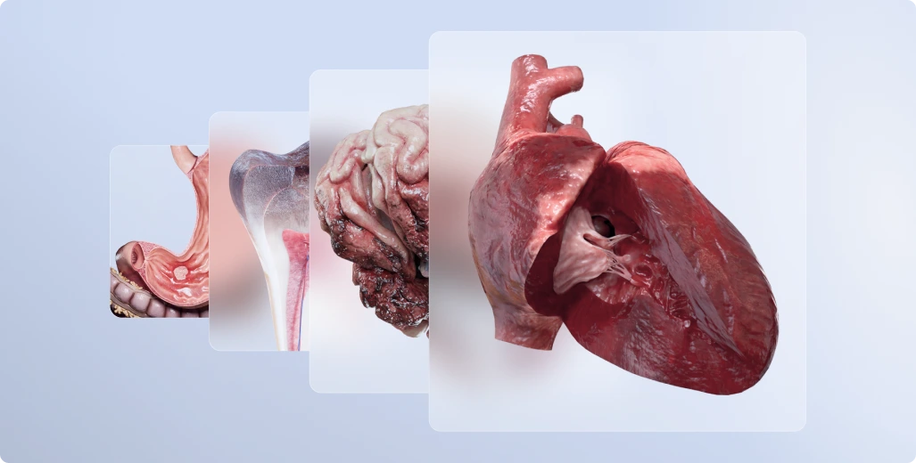
Digital 3D models: virtual, three-dimensional representations of tissues, organs, or systems created with advanced imaging techniques and computational reconstruction. These 3D pathology models provide a detailed view of damage caused by diseases.
One of the key advantages of such models is their versatility. They can be turned into educational videos and 3D animations to demonstrate disease progression or treatment effects or used in AR/VR environments (although this is not yet standard practice and requires specific hardware/software and training).
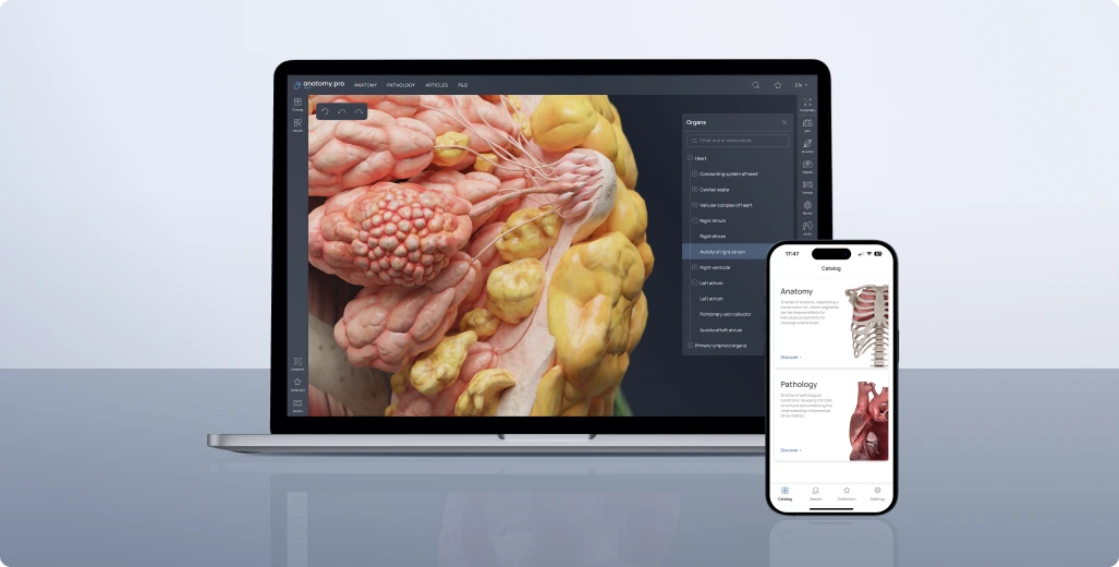
However, the quality of a 3D model is directly dependent on the resolution of the source imaging data used for the model creation. VOKA 3D Anatomy and Pathology offers the most comprehensive experience for exploring digital 3D models. Its high-quality anatomical and pathological models can be easily integrated into medical training, diagnostics, and treatment planning. -
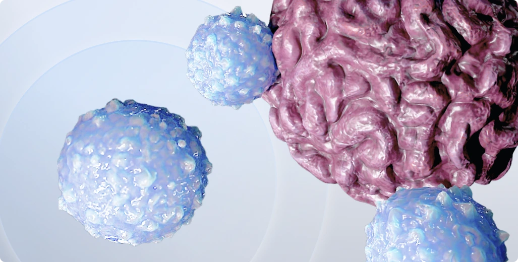
3D in vitro models: laboratory-grown cultures that mimic actual human cells and tissues. Examples include organoids, organ-like structures grown from stem cells (e.g., liver organoids or brain organoids), and spheroids, compact clusters of cells that can simulate tumors or affected tissues.
It's important to note that while valuable, these models are simplified representations and may not fully replicate the complex in vivo environment, such as cell-cell and cell-matrix interactions, vascularization, and immune responses. Digital models, in turn, are flexible enough to simulate a wide variety of structures, from whole organs down to individual cells and even subcellular components. -
3D-printed models: physical replicas of tissues and organs created using the 3D printing technology. Digital 3D models serve as blueprints guiding the printing process. Non-biological 3D-printed models, made from materials like plastic or resin, are widely used in medical education, patient communication, and research. These models help explain complex anatomy, simulate surgical procedures, and test medical devices in R&D settings.
Bioprinted models, on the other hand, use bioinks containing living cells to create functional, tissue-like structures. These are primarily used in advanced medical research to test drug responses, study disease mechanisms, or explore regenerative medicine. Some challenges of 3D bioprinting include achieving high resolution for intricate microstructures and successfully vascularizing larger tissues.
Limitations of 2D images in visualizing pathologies
While traditional 2D images remain a gold standard for many diagnostic purposes, particularly at the cellular level, more complex cases require better dimensional depth. Here are some limitations of the 2D approach:
Loss of spatial context
Since 2D images offer only a flat view, it’s difficult to see the true spatial relationships between cells, tissues, and organs. In terms of pathology research, this limitation can obscure how diseases progress, how affected cells interact, or how structural abnormalities influence surrounding tissues.
A vivid example of this limitation is the study of the extracellular matrix (ECM), a critical component of tissues that provides physical and biochemical support to cells. In 2D images, the ECM looks overly simple, which hides its complex network. Since the ECM influences processes like cell migration, adhesion, and tissue repair, missing these spatial details can limit our understanding of oncology cases.
Limited representation of complex structures
When viewed in 2D, biological structures like blood vessels, tumors, and neural networks lose their true shape and complexity. For example, tumors often grow in unpredictable, irregular patterns as they spread into nearby tissues and create intricate connections with blood vessels. A single 2D image captures only one side of this structure. As such, a limited view makes it difficult to fully understand the tumor’s exact shape and how far it has spread.
For doctors and researchers, assessing a tumor's growth pattern and spread requires multiple 2D images, which is less efficient and comprehensive than a 3D representation.
Oversimplification of tissue heterogeneity
Pathological tissues are highly heterogeneous: they comprise different cell types, structures, and microenvironments. However, when analyzed through 2D images, this complexity is oversimplified. As a result, doctors can deal with an inaccurate representation of the tissue’s behavior.
Speaking of tumors again, such pathologies can contain regions with active cell division, areas of dead cells, and zones with dense blood vessel growth. In a 2D image, these diverse regions can be easily overlooked or misinterpreted, as the complete picture of the tumor’s heterogeneity isn’t visible. This can affect critical research and clinical decisions, such as determining how aggressive a tumor is or how well it might respond to treatment.
How 3D pathology models enhance disease comprehension
3D models offer researchers and practitioners a more detailed understanding of diseases. Here’s how these models are changing the way we study and comprehend pathologies:
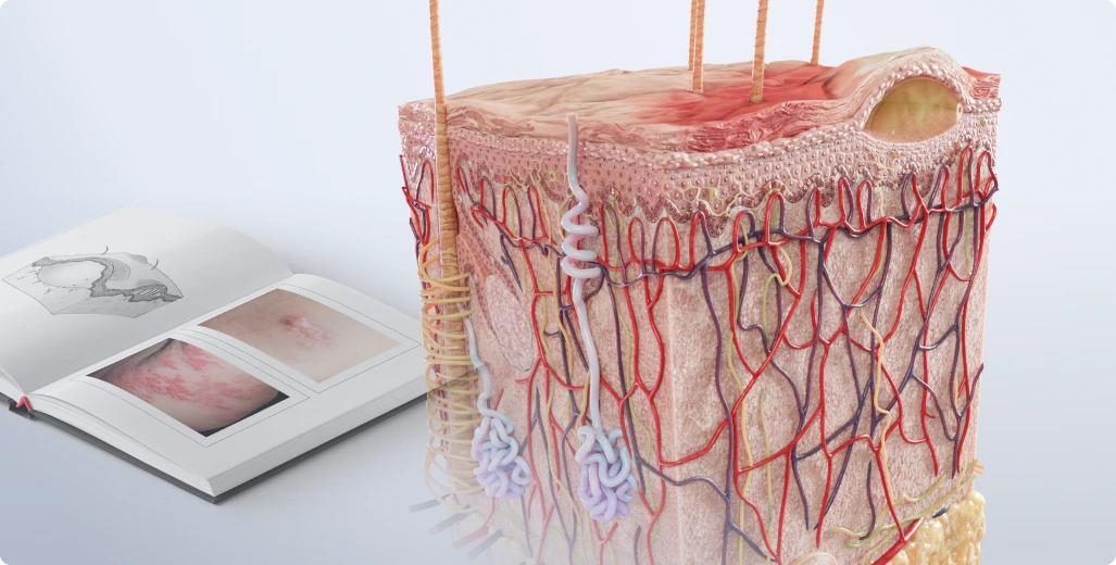
Improved visualization of affected tissues and organs
3D pathology models let us explore damaged systems from multiple angles and layers. Unlike traditional 2D methods, they offer a complete picture, which makes understanding the impact of various diseases easier.
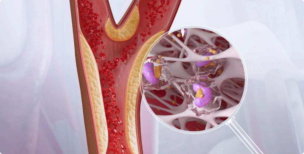
Let’s take the study of cardiovascular diseases as an example.
With digital 3D models, like those in VOKA 3D Anatomy and Pathology, healthcare professionals and students can visualize blood flow through vessels and pinpoint the effects of blockages, which is why they are becoming essential interactive medical training solutions. For example, a 3D pathology model can demonstrate how a specific stenosis (narrowing) in a coronary artery affects blood flow to a particular region of the heart muscle.
In vitro models can simulate plaque buildup, allowing researchers to study the dynamics of its development and its impact on vessel function. Specifically, researchers can observe how different shear stresses within the vessel affect plaque formation and stability.
Also, 3D printing can create physical replicas of damaged heart valves, and surgeons can use those to practice repairs and plan individualized surgical approaches. This physical preparation is often complemented by detailed surgical procedure animations, which help the surgical team clearly visualize every step of the intervention before entering the operating room.
Simplified study of cell-ECM interactions
The extracellular matrix (ECM) is not merely a structural scaffold; it actively influences cell behavior. Understanding cell-ECM interactions is crucial for comprehending tissue repair mechanisms, disease progression (such as cancer metastasis), and the development of targeted therapies. 2D methods oversimplify these dynamics and hinder effective research.
In contrast, 3D pathology models create realistic, three-dimensional environments where researchers can observe and manipulate key factors like matrix stiffness, ligand density, and porosity. For example, a 3D model of a tumor can reveal how changes in ECM stiffness influence cancer cell migration and invasion. By replicating in vivo conditions, 3D models provide a more accurate platform for studying these crucial interactions.
Dynamic simulations of disease dynamics and behavior
3D models can simulate disease progression in a controlled environment, revealing critical stages that might be missed in static images. Examples include:
-
Tumor growth and metastasis: digital 3D models can be used to create 3D animations of tumor growth, visualizing how cancer cells proliferate, invade surrounding tissues, and potentially metastasize to distant sites. These simulations can include data from patient scans to create personalized 3D medical models of tumor development. In vitro 3D tumor models can be used to study the effects of different drugs or therapies on tumor growth and invasion in real-time.
-
Infectious disease spread: 3D models can simulate the spread of infections, showing how pathogens interact with host tissues and the immune system. This can help researchers understand the dynamics of infection and develop strategies to control its spread.
Drug response testing
3D digital models offer a foolproof way to test how drugs interact with tissues in a realistic environment.
They can be used to create visualizations of drug diffusion, absorption, and resistance within tissues. For example, a 3D model of the liver can be used to simulate how a drug is metabolized and distributed throughout the organ.
In vitro 3D models allow researchers to observe real-time cellular responses to drugs, such as the effects of chemotherapy on cancer cells or the impact of a new drug on healthy tissues. These models can help optimize dosages and drug combinations for safer and more effective treatments.
Additionally, the 3D models can be used along with MoA animations that will demonstrate the drug response mechanisms in movement. Such a combined approach provides additional aid in understanding the optimal drug dosages and effects of drug combinations.
Real-life applications of 3D pathology models in medical fields
3D pathology models have a wide range of applications across medical fields, providing new insights that were once out of reach. Here are some real-life examples of how different types of 3D pathology models are making a difference:
Oncological diseases
3D models have significantly advanced cancer diagnostics. A study published in JAMA Network Open highlights the use of 3D medical models in oncology, specifically in planning robotic-assisted laparoscopic radical prostatectomy (RALRP) for prostate cancer patients.
These digital models were created using preoperative imaging data, such as MRI. As a result, specialists were able to create highly detailed and accurate 3D representations of the prostate, surrounding tissues, and the tumor itself.
Using these 3D models, surgeons could better visualize the spatial relationships between the tumor and critical structures such as nerves and blood vessels. The models were even made accessible via a mobile app on the surgeons' smartphones, which they could consult before and during surgery. In addition to planning, clinics often utilize specialized oncology animation services to simplify these complex case details for patient education.
Neurological disorders
One of the most promising applications of 3D models is the study of pathologies such as autism, Alzheimer's disease, and Parkinson's disease (PD). According to the Journal of Biomedical Science research, in vitro 3D brain organoids may be the groundbreaking tools for understanding and treating neurological disorders. These organoids were derived from human induced pluripotent stem cells (hiPSCs) and engineered to mimic the structure and function of the human brain.
The in vitro models allowed researchers to observe how abnormalities in brain development contribute to neurological disorders. They have also served as a platform for testing potential therapies, allowing scientists to evaluate drug effects in a controlled environment.
Cardiovascular diseases
3D pathology models and custom cardiology animations are widely used in the diagnosis and treatment of complex cardiovascular diseases. Such models are created using imaging techniques such as CT and MRI scans, which are then transformed into patient-specific digital 3D models and then printed. A study by Micromachines explains how 3D models provided surgeons with an accurate representation of the patient's heart anatomy, including defects, blood vessels, and structural abnormalities.
One notable application detailed in the study is the use of 3D-printed heart models for planning procedures such as aortic valve replacement and repair of congenital heart defects. These models allowed surgeons to visualize and simulate the procedure before entering the operating room, reducing surgical risks and improving outcomes. The tactile nature of the 3D printed models makes the treatment process more personalized.
Infectious diseases
3D models have shown great potential in improving diagnostic accuracy and patient care in infectious disease management. According to BMJ Case Reports, during the early stages of the COVID-19 pandemic, clinicians used 3D reconstructions of lung CT scans to better understand the extent and distribution of lung damage caused by the virus. These 3D models provided a detailed visualization of infected areas, enabling accurate measurement of disease progression and severity, even in cases where traditional diagnostic methods such as RT-PCR were inconclusive.
Healthcare professionals were able to tailor interventions such as oxygen therapy or ventilator settings to each patient's specific needs by visualizing the exact regions of the lung affected by COVID-19. In addition, the 3D pathology models were used for patient education and communication, helping patients and their families understand the severity of the disease, evaluate proposed treatment plans, and provide informed consent.
Wrapping up
3D pathology models have changed the way medical professionals study, diagnose, and treat disease. From oncology to infectious diseases, 3D models have proven to be versatile tools that improve medical research and clinical outcomes.
Innovative anatomy and pathology resources, such as VOKA 3D Anatomy and Pathology continue this approach by providing ready-to-use pathology models for medical professionals and educators. In addition, the VOKA team can develop custom 3D pathology models tailored to individual patient conditions if you need patient-specific visuals for training or patient communication. With tools like these, the possibilities for improving healthcare are endless.
Make VOKA your preferred source
See more VOKA articles in Google Search
Share
Table of contents
Thank you for your comment!
Your comment has been submitted for moderation and will be published soon. We'll email you once it’s live.