
3D cell structure animation: enhancing pharma research and drug development
Share:
Copy link
Share via Telegram
Share on Facebook
Share on LinkedIn
Share via WhatsApp
Share on X
Share via Email
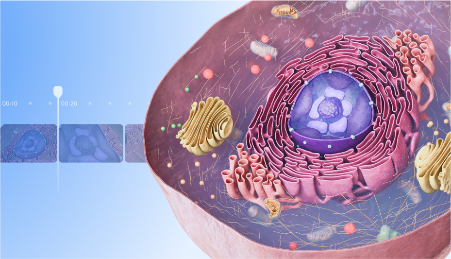
Table of contents
What happens inside the human body during illness? Beyond the sore throat or runny nose, a complex chain of events unfolds, and most of it’s completely invisible to the naked eye. As soon as bacteria or viruses enter the body, the immune system responds. Signals are sent to raise body temperature, blood vessels dilate to let in white blood cells, and energy is redirected to fuel the body’s defenses. The fatigue and discomfort that follow are signs of a full-scale response happening on a microscopic level.
At the center of it all is the cell. Every symptom, every reaction, and every healing process begins there. But these microscopic events are far too small to observe directly — and without visibility, understanding the root causes of diseases and designing effective therapies becomes much harder. That’s where cell structure animation comes in: by making the unseen seen, it can help researchers uncover potential pathways for treatment.
Why is understanding cell structure important?
The better we can see and grasp what happens inside the cell, the more precise and effective treatments will be. Eukaryotic cells, with their complex structures and processes, are at the center of most diseases and drug interactions. Understanding cells and their key features is critical for uncovering disease mechanisms and determining how drugs interact with specific cellular components. In the following section, we’ll explore the key organelles and their functions, highlighting their importance in biomedical research and early-stage drug development.
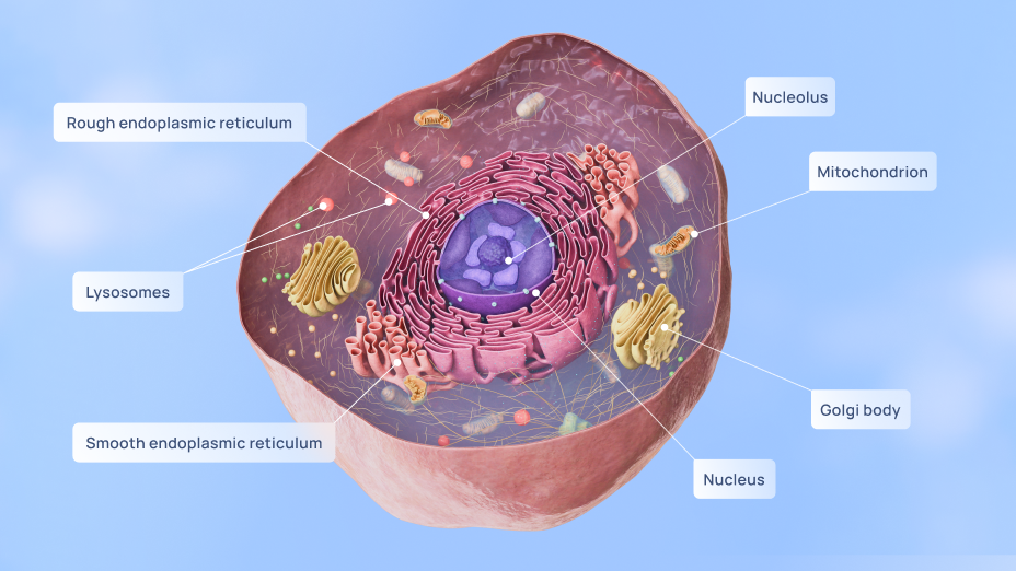
The cell membrane: gatekeeper and drug target
The cell membrane acts as the gatekeeper of the cell — regulating what enters and exits. Because many drugs must cross the lipid bilayer or interact with membrane proteins, it’s a critical focus for pharmaceutical research. Visualizing this barrier helps teams model drug absorption, anticipate intracellular transport, and understand potential resistance mechanisms.
Mitochondria: energy and early warning signals
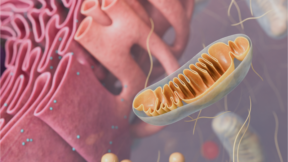
Beyond powering the cell, mitochondria regulate metabolism, apoptosis, and stress responses. Their dysfunction is linked to conditions ranging from neurodegeneration to cancer. By studying how mitochondria respond to new compounds, researchers can better assess a drug’s impact on cellular health, making them vital for early efficacy and toxicity screening.
The endoplasmic reticulum: the critical role of protein folding
The endoplasmic reticulum is where proteins are built and folded. In many diseases — including cystic fibrosis, diabetes, and cancer — misfolded proteins trigger cellular stress. For drug developers, understanding how the endoplasmic reticulum responds to stresses like protein misfolding or calcium imbalance is essential for effectively targeting these pathways.
The nucleus: command center for genetic targets
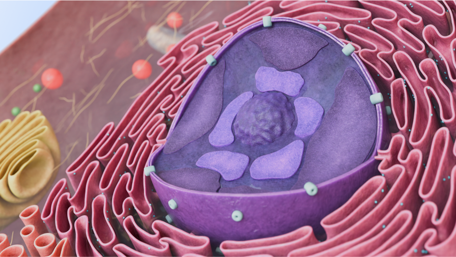
The nucleus contains DNA and coordinates gene expression. Many modern drugs, including gene therapies and cancer treatments, target this control hub. Understanding how genetic material is organized, how transcription is triggered, and how therapies interact with DNA-level processes is crucial for developing more precise and effective treatments targeting genetic disorders and cancers.
Cytoskeleton: not just for structure
The cytoskeleton gives shape to the eukaryotic cell, but it also drives movement, intracellular transport, and cell division — key processes in tumor growth and metastasis. Targeting cytoskeletal elements can disrupt disease progression, making it a growing area of drug research. Detailed animations help researchers explore these dynamics and test hypotheses in silico before lab trials begin.
Key obstacles in pharma research & drug development
To develop effective therapies, researchers must navigate biological variability, unpredictable responses, and high failure rates within the nanoscopic structure of cells. Below are some of the key challenges the industry continues to face in studying how cellular and subcellular structures behave and interact during the drug development process.

High failure rates in preclinical and clinical stages
Most drug candidates never make it past early testing. In fact, the probability of a drug making it from Phase I to approval is only 7.9%, according to BIO's Clinical Development Success Rates report. Without a clearer picture of what’s happening at the cellular level during a cell's interaction with the drug candidate, it remains difficult to predict outcomes and interpret scientific experimental data.
Challenges in validating molecular targets in drug development
It’s debatable that targeting a specific molecule will lead to therapeutic benefits in humans before a drug is developed. This is because lab tests, animal models, and human diseases can behave differently, making it hard to accurately validate whether the target will be effective in humans. These biological differences complicate the process of confirming that a target will have the desired therapeutic effect across different models and in humans, according to research published in PMC.
Communication gaps across research teams
Pharma R&D often brings together biologists, chemists, data scientists, and clinicians. But communicating complex cellular data across disciplines can be challenging. For example, biologists and data scientists may interpret the same data differently. Without a shared conceptual or visual framework, critical nuances may be missed, leading to misalignment and slowed collaboration across the team.
How medical animation enhances cell structure, pharma research & drug development
Pharmaceutical research is under pressure to move faster, cut costs, and improve accuracy — all without compromising safety. But there’s a powerful tool emerging in response. Cell structure animation. By visualizing cellular and subcellular activity in high resolution, animation helps researchers uncover how drug candidates behave, validate earlier, and more. Let’s explore the core benefits of medical animation now.
Revealing cellular resistance and drug interactions
In diseases like cancer, cells can stop responding to the treatment, making it less effective, as BMC research reports. 3D medical animations, such as cell membrane structure and receptor interaction animations, can visualize how resistance mechanisms emerge at the cellular level, whether through mutations or changes in membrane permeability. Early visibility helps teams tweak compounds before costly trial phases.
Accurate depictions of disease mechanisms
Researchers can visualize through MoD animations how cells behave during disease progression — whether it's cancer cells multiplying, infected cells responding to a virus, or mutated proteins causing dysfunction. These clear depictions allow pharma teams to explore how specific treatments can target and reverse these processes, potentially increasing the chances of developing effective therapies.
Enhancing safety assessments and toxicity studies
Understanding off-target or long-term effects is critical in drug development, especially when dealing with diseases like cancer or neurodegenerative disorders. For instance, cancer drugs might impact not only the tumor cells but also healthy cells, and using 3D cell structure animation can visually simulate the progression of cellular changes over time, helping researchers better understand how drugs interact with different cell types.
Streamlining cross-disciplinary collaboration
Pharma teams span biology, chemistry, data science, and clinical practice — and often struggle to align around abstract findings. High-fidelity cellular animations give all stakeholders a shared visual language. This streamlines collaboration, reduces misinterpretation, and speeds up decisions across departments and specialisms.
Visualizing complex biology for smarter research decisions
Above all else, having a clear understanding is everything in medical research and drug development – even more so when dealing with complex biological systems. VOKA’s 3D animation services turn intricate cellular and molecular processes into clear, dynamic visuals that, ultimately, drive better research outcomes.
With anatomically precise modelling and scientific accuracy, VOKA helps researchers and pharma teams visualize everything from drug-receptor interactions to disease progression and therapeutic mechanisms – all at the cellular, molecular, and systemic levels. These animations go far beyond static diagrams, making it easier to spot key pathways, explore treatment effects, and communicate findings across disciplines.
By making complexity accessible, VOKA animations help identify potential risks earlier, improve cross-team alignment, and support smarter, faster decision-making throughout the development lifecycle.
Final thoughts
3D cell structure animation provides clear, detailed visualizations that help researchers better understand complex biological processes. By providing a dynamic, accurate visual of cellular mechanisms and disease progression, it helps researchers see exactly how drug molecules interact with cells. This deeper insight is invaluable for identifying potential problems early in the development process, such as how a treatment might influence a disease or how resistance mechanisms may arise. The ability to visually track these processes improves collaboration across research teams, supports clearer communication, and enables a more informed approach to decision-making.
Share
Table of contents
Thank you for your comment!
Your comment has been submitted for moderation and will be published soon. We'll email you once it’s live.