
Volume rendering
visualizes the scan as a 3D image
Surface reconstruction
converts selected anatomy into 3D geometry
Automation
creates a starting point, not a presentation-ready model
Human refinement
cleans the geometry for further visualization
We reconstruct CT and MRI DICOM data into anatomically accurate 3D models through manual segmentation and in-house clinical review. The resulting 3D assets can be used in medical animations, web-based viewers, and interactive XR experiences.
Patient-derived
Built from the original CT or MRI study
Clinician-reviewed
Reviewed by 20+ in-house clinicians
Anatomically correct
Hand-cleaned geometry with structures separated and named
CT and MRI scans contain volumetric data captured as a series of 2D slices. 3D reconstruction turns this data into surface geometry, creating a patient-specific 3D model from selected anatomical structures.
visualizes the scan as a 3D image
converts selected anatomy into 3D geometry
creates a starting point, not a presentation-ready model
cleans the geometry for further visualization



process
From DICOM to a usable 3D asset
process
The same CT/MRI reconstruction can be turned into different visual assets without rebuilding the anatomy from scratch.
Animations for congress presentations, MoA/MoD storytelling, HCP education, and patient communication.
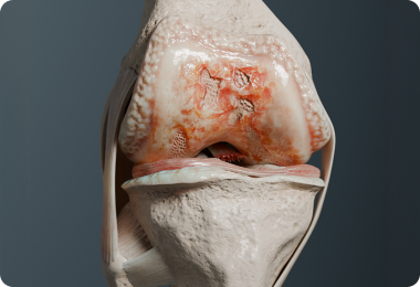
Clean, watertight geometry for 3D printing or CAD workflows in STL, OBJ, 3MF, STEP, and other formats by request.

High-fidelity visuals for publications, posters, presentations, and promotional materials for industry events.
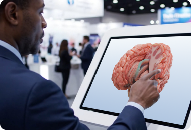
Anatomy visualization presented as an interactive 3D viewer for websites and educational modules.
Patient-specific 3D scenes designed for immersive VR and AR experiences, from educational modules to training environments.

Vessels & aorta
Aortic and vascular models, including the Circle of Willis, for HCP education.
Heart & coronary arteries
Coronary anatomy from CT angiography for education and scientific content.
Teeth & maxillofacial structures
High-resolution CBCT models for dental products, treatment planning, and research.
Bones, joints & trauma
Patient-specific models of the shoulder, pelvis, and Hill-Sachs lesions.
Face & craniofacial structures
Facial models capturing bone, soft tissue, and spatial relationships.
Brain & neurovasculature
Brain and neurovascular models for exploring complex anatomical relationships.
Abdomen & urinary tract
Urinary and abdominal structures, including CT urogram models.
CT & CTA
Reconstruction of bone, soft tissue, and vascular anatomy, including contrast-enhanced CT angiography.
MRI
3D reconstruction from MRI data with detailed representation of tissue boundaries and internal structures.
CBCT
Detailed 3D reconstruction for dental, maxillofacial, and other high-resolution applications.
Micro-CT & research imaging
High-resolution 3D reconstruction for research applications and specialized imaging datasets.
Our 3D reconstruction process combines expert 3D work with clinical review at every stage
| Key criteria | DIY tools | 3D studio/freelancer | Imaging bureau | VOKA |
|---|---|---|---|---|
| Works from original DICOM | Yes, manually | Usually not | ||
| Manual segmentation | Done by your team | |||
| Clinical anatomical review | Geometry only | |||
| Complex anatomy & vascular reconstruction | Possible, but labor-intensive | Limited | ||
| Your team’s involvement | High | Medium | Low | Low |
VOKA combines medical expertise and efficient production to deliver experiences for real-world communication needs.

Deep medical proficiency
Our development team and in-house medical specialists ensure simulations are anatomically and procedurally accurate.

Rapid deployment & cost-efficiency
We leverage our extensive library of 3D models to quickly and cost-effectively build interactive medical scenes.
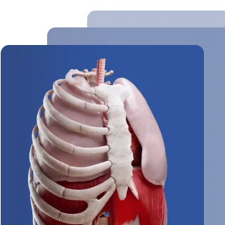

Precision in 3D content
We ensure that every anatomical structure and pathology we create meets the highest standards of medical precision.

Tailored & scalable solutions
Beyond our ready-made assets, we offer custom development to align with narratives of any complexity.
Recognized for excellence in healthcare animation






Explain MoA, disease mechanisms, and clinical concepts through anatomy reconstructed from real patient imaging for HCP education and clinical communication.

Assess device concepts, anatomical fit, and spatial relationships using detailed 3D reconstructions of real anatomy.

Strengthen scientific and medical communications with accurate, patient-derived anatomical visuals for campaigns, publications, and congresses.

Standardize anatomical models and visual datasets derived from medical imaging for research, preclinical studies, and publications.

Prepare case discussions, surgical training, and patient communication with detailed reconstructions of complex anatomy and pathology.

Connect theoretical anatomy with real imaging data through detailed 3D models of complex structures and pathology.

Book a call or fill out the form and we’ll get back to you once we’ve processed your request.
You can contact us using:
FAQ
What is a 3D reconstruction CT scan?
A 3D reconstruction turns CT scan data into a three-dimensional model of the anatomy. It uses the original image slices to create a detailed surface representation of bones, organs, vessels, or other structures.
What is the difference between a normal CT scan and a 3D CT scan?
A CT scan produces a series of cross-sectional images. A 3D CT reconstruction uses those images to create a three-dimensional model that can be viewed, edited, rendered, or used in other visual applications.
What does CT reconstruction mean?
Computed tomography reconstruction is the process of turning CT image data into a 3D representation of anatomical structures. It can range from automated volume rendering to a manually refined 3D model.
What are the advantages of 3D CT reconstruction?
A 3D model makes complex anatomy easier to understand and communicate. It can also serve as the basis for animation, 3D rendering, and interactive VR/AR experiences.
What files do you need from us?
We work with DICOM, NIfTI, and NRRD files. The quality of the reconstruction also depends on factors such as slice thickness, image resolution, and contrast phase.
What slice thickness do you need for a reliable reconstruction?
There is no single ideal thickness for every case. Thinner slices generally provide more detail, but the right parameters depend on the anatomy, imaging protocol, and structures being reconstructed.
Who reviews the model, and what does the review cover?
Each model is reviewed by our in-house clinicians. The review covers anatomical structures, boundaries, separation, naming, and overall consistency with the imaging data.
How long does a reconstruction project take?
The timeline depends on the complexity of the anatomy and the level of refinement required. It typically includes study assessment, segmentation, anatomical cleanup, clinical review, and final delivery.
Did not find an answer to your question?
You can write to us by email and our manager will answer all your questions: