
Digital anatomy: reshaping the study of the human body
Share:
Copy link
Share via Telegram
Share on Facebook
Share on LinkedIn
Share via WhatsApp
Share on X
Share via Email
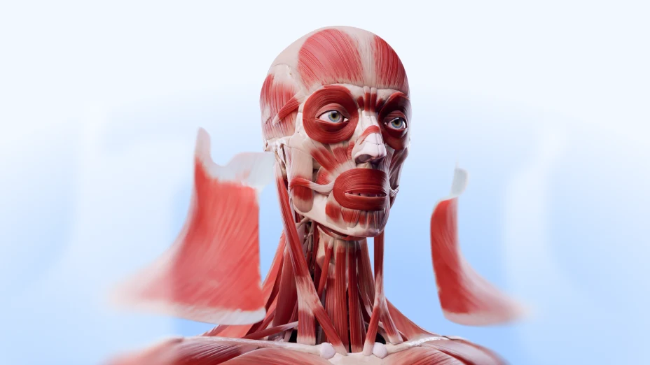
Table of contents
Learning human anatomy has always been challenging. The human body is made up of hundreds of structures, layers, and systems, all interconnected in ways that are difficult to visualize.
For educators, this complexity creates a teaching hurdle. Traditional diagrams and textbooks often fall short, leaving students struggling to translate 2D images into a 3D understanding of the body. What’s needed is a way to explore anatomy that’s both accurate and immersive.
That’s exactly where digital anatomy comes in. By combining interactive 3D models with advanced visualization tools, digital anatomy makes it easier to teach, learn, and truly grasp how the human body works.
In this article, we’ll explore the definition of digital anatomy, explore how it’s reshaping medical education, and highlight the opportunities it creates for both teachers and students.
What is digital anatomy?
The term digital anatomy refers to the use of interactive, three-dimensional modeling and visualization of human anatomy through advanced technologies.

Unlike traditional anatomy, which relies heavily on physical dissection and two-dimensional illustrations, digital anatomy offers a dynamic and immersive way to explore the human body.
Key technologies involved in digital anatomy include:
-
3D modeling,
-
Virtual reality (VR),
-
Augmented reality (AR),
-
Mixed reality (MR),
-
3D printing, among others.
The methods and technologies of digital anatomy are increasingly being adopted in medical education, helping students and professionals explore complex structures more intuitively.
Digital anatomy also plays a crucial role in surgical planning, allowing surgeons to rehearse procedures and visualize patient-specific anatomy. Additionally, it supports research by providing accurate, manipulable models and enhances simulation training for medical scenarios.
Simply put, if traditional anatomy is about dissection in the lab, the digital anatomy meaning refers to exploration in a 3D space.
Digital anatomy term: evolution and context
Digital anatomy is the result of years of innovation in medical imaging and education technology. As healthcare entered the digital age, anatomy education began to shift too, from textbooks and cadavers to tablets, headsets, and 3D models.
The digital anatomy term itself is relatively new but growing fast in popularity. It fits within the broader movement toward digital transformation in healthcare and education, often called Health 4.0.
From sketches to simulations
Anatomy has always been a visual science: first taught through hand-drawn atlases, later with plastic models, cadaver labs, and radiological imaging. Let’s take a quick look back at its evolution:
Ancient times: Early physicians like Hippocrates and Galen relied on verbal descriptions and basic sketches to understand the body.
16th century: Andreas Vesalius revolutionized anatomy with De humani corporis fabrica (1543), a detailed atlas featuring hand-drawn, highly accurate anatomical illustrations based on human dissections.
18th–19th centuries: Plastic and wax models became popular teaching tools, offering 3D representations without the need for cadavers. At the same time, cadaver dissection labs remained the gold standard for hands-on learning.
20th century: The advent of medical imaging transformed anatomy education. X-rays in the early 1900s allowed visualization of bones inside the living body. Later, advances like CT in the 1970s and MRI in the 1980s provided detailed cross-sectional and soft tissue images.
Late 20th to early 21st century: These imaging breakthroughs were digitized, paving the way for 3D reconstructions. Computer graphics technology matured, enabling interactive models that students could manipulate on screens.
Today: Digital anatomy integrates high-resolution 3D models, AR/VR, and more, to make anatomy learning immersive, interactive, and accessible worldwide.
Part of a bigger shift: Health 4.0
The digital human anatomy definition goes beyond virtual models of the body — it represents a core element of the Health 4.0 movement, where healthcare and technology converge. Just as engineers use digital twins to test and refine complex systems, medicine now has its own digital twin of the human body.
This shift is already transforming education, research, and clinical practice. Students can now dissect a lifelike virtual cadaver without the limitations of physical specimens. Surgeons can rehearse complex procedures on patient-specific digital models before entering the operating room. Researchers can simulate how tissues respond to stress or treatment, accelerating discovery while reducing risk.
Why it matters now
Modern learners want flexible, tech-integrated education. Many students find traditional cadaver dissection overwhelming, logistically difficult, or even emotionally uncomfortable. At the same time, schools face a shortage of cadavers and trained anatomy educators.
Digital anatomy offers a scalable, accessible, and adaptable solution to any setting. It’s not just a trend. It’s the evolution of anatomy as both a discipline and a teaching method, shaped by the digital transformation of medicine.
Tools and technologies powering digital anatomy
When we define digital anatomy, we mean the use of advanced tools and technologies that transform traditional anatomical study. The key technologies powering digital anatomy include:
3D modeling
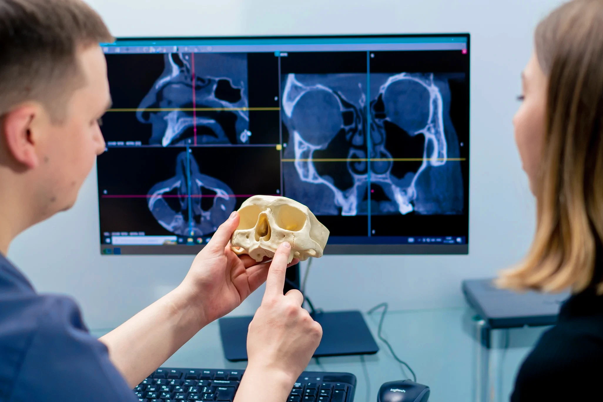
3D modeling is the foundation of digital anatomy. It involves creating accurate, high-resolution digital representations of anatomical structures using specialized software. These models are often developed from medical imaging data such as CT scans, MRI, or microscopy, allowing for precise anatomical detail and realistic visualization.
A wide variety of 3D anatomical atlases and platforms have been developed to facilitate this immersive learning. A great example is VOKA 3D Anatomy and Pathology, which stands out for combining detailed anatomical structures with corresponding pathological changes. Users can rotate, zoom, dissect, and isolate parts of the model, providing a comprehensive understanding of spatial relationships within the human body. This dynamic manipulation offers a much richer experience compared to traditional static images or physical specimens.
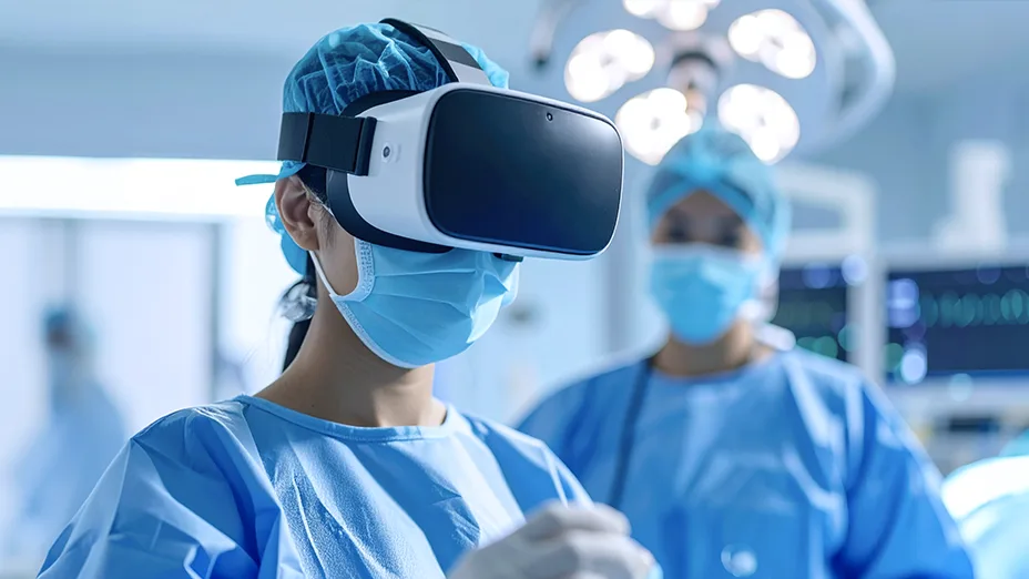
Virtual reality (VR)
Virtual reality immerses users in a fully digital, three-dimensional environment where they can interact with anatomical models in real time. By wearing VR headsets, students and clinicians can explore the human body from the inside out, virtually “walking through” organs or systems, simulating procedures, and gaining a deeper spatial awareness.
Beyond education, VR plays a pivotal role in surgical simulation and training. Surgeons can rehearse complex procedures in a risk-free virtual environment, practicing techniques and anticipating challenges before operating on actual patients.
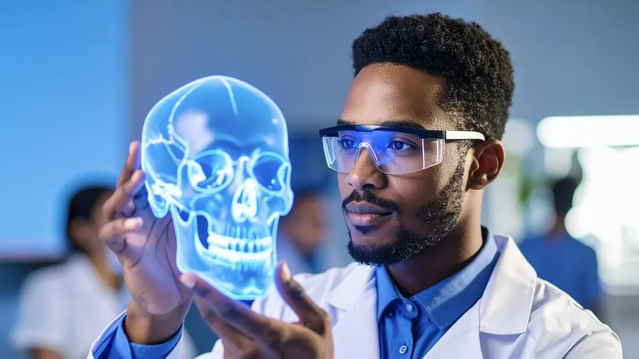
Augmented reality (AR)
Augmented reality overlays digital anatomical information onto the real world through devices like smartphones, tablets, or AR glasses. This technology allows users to visualize and interact with 3D anatomical structures superimposed on physical objects or even directly on a patient’s body. By blending virtual elements with the physical environment, AR provides an intuitive way to study anatomy in context.
In medical education, it offers students the ability to explore anatomy hands-on while maintaining interaction with their surroundings. For patient communication, AR helps clinicians explain complex conditions or surgical plans in a clear, visual manner, improving patient engagement and informed consent.
Mixed reality (MR)
Mixed Reality combines elements of both VR and AR, blending physical and virtual environments to create interactive, context-aware experiences. MR devices, such as Microsoft HoloLens, enable users to manipulate digital anatomy models within their real surroundings while maintaining awareness of the physical world. This fusion enhances collaborative learning and surgical planning by allowing multiple users to interact with the same digital anatomy in a shared space.
For institutions and healthcare companies that want to move beyond ready-made anatomy platforms, custom immersive applications can help adapt digital anatomy to specific educational, clinical, or product needs. VOKA’s VR & AR development service supports the creation of tailored healthcare experiences, from interactive anatomy training and surgical simulation to patient education and medical product demonstrations.
3D printing
3D printing converts digital anatomical models into tangible physical objects. Using various printing materials, complex anatomical structures can be recreated with high fidelity, offering hands-on learning tools and patient-specific surgical guides. These physical models are invaluable for preoperative planning and education, allowing tactile exploration and rehearsal that complement virtual experiences.
In addition, 3D-printed models are increasingly used for marketing and demonstration purposes by medical device companies and healthcare institutions, showcasing implants, prosthetics, or surgical innovations in a visually compelling way.
Transition to digital anatomy: opportunities and barriers
As the world steadily moves toward digital solutions in education and healthcare, anatomy is no exception. However, the transition from traditional methods comes with its own set of opportunities and challenges. Let’s explore them in detail:
1. Economic and resource efficiency
One of the strongest arguments for adopting digital anatomy is its potential to optimize costs and resources over time. Traditional cadaver-based teaching remains a cornerstone of medical education, but it involves significant ongoing investment in specimen procurement, storage facilities, staff, and compliance with health and safety regulations.
Digital platforms do not eliminate the need for cadaver labs but can complement them by reducing the pressure on resources, offering scalable access, and supporting remote or hybrid learning environments. While initial setup costs for digital tools can be high, their value grows over time when used alongside traditional methods.
Opportunities
-
Long-term cost efficiency by reducing recurring expenses for cadavers, storage, maintenance, and safety procedures.
-
Low ongoing maintenance costs once hardware and software are in place.
-
Potential for scalability, as digital resources can often be shared among larger groups of students (note that costs may still rise with licensing fees).
-
Remote accessibility allows for flexible, time-efficient learning and reduces the need for physical space.
Barriers
-
High initial setup costs for VR/AR hardware, computers, and specialized software licenses.
-
Infrastructure compatibility issues, particularly in cadaver-based labs with older equipment or systems.
-
Ongoing upgrade expenses due to rapid technological changes.
2. Educational transformation and learner experience

Digital anatomy transforms how students engage with complex anatomical concepts by merging surface, regional, and internal anatomy into one interactive environment. It enables virtual dissection, integrates multiple disciplines, such as anatomy, physiology, and pathology, and allows side-by-side comparison with medical imaging.
These capabilities can enhance comprehension, retention, and clinical reasoning while supporting both self-directed and collaborative learning. However, limitations such as reduced tactile feedback, incomplete representation of anatomical variations, and varying digital literacy levels among learners and instructors can hinder its full impact.
Opportunities
-
Integration of multiple disciplines (e.g., anatomy, physiology, pathology) in a single interactive platform.
-
Ability to combine gross and microscopic anatomy with medical imaging for a richer context.
-
Consistent learner satisfaction has been reported across various studies.
-
Augmented and virtual reality enhancements are making experiences more immersive and lifelike.
Barriers
-
Limited tactile feedback, which can be important for developing surgical and procedural skills (unless addressed by VR and haptic solutions).
-
Variable digital competencies among instructors and students are affecting engagement and learning outcomes.
-
Limited exposure to certain emotional or professional aspects of traditional anatomy education, such as confronting death in cadaver labs.
3. Technology integration
The success of digital anatomy depends heavily on how seamlessly it integrates with existing educational and clinical infrastructures. Effective integration ensures that VR, AR, MR, and other digital tools complement rather than disrupt established curricula and workflows.
When supported by compatible systems, reliable networks, and proper training, these technologies can enhance the learning experience and expand access. However, incompatibility with older facilities, the need for frequent updates, and the pace of technological change can pose significant challenges to sustained use.
Opportunities
-
Compatibility with remote learning platforms and hybrid teaching models.
-
Cloud-based access enables resource sharing across multiple locations.
-
Integration with hospital-based skills labs for both academic and clinical applications.
-
Potential to link with emerging technologies such as AI-assisted anatomy labeling or real-time pathology mapping.
Barriers
-
Infrastructure limitations in institutions with older or incompatible equipment.
-
High upgrade frequency due to fast-moving VR/AR hardware and software development.
-
Potential technical disruptions from network or software instability.
-
Need for specialized IT support and staff training to maintain and operate systems.
-
Risk of fragmented learning experiences if tools are poorly integrated into the curriculum.
4. Clinical and research applications
Digital anatomy extends far beyond the classroom, offering valuable tools for clinical practice and medical research. In clinical settings, it supports surgical planning, patient education, and intraoperative guidance by providing highly detailed, manipulable 3D models. In research, these tools enable advanced visualization, data integration, and cross-institution collaboration, fostering innovation in personalized medicine and procedural development. While the potential is significant, successful adoption depends on technological maturity, interdisciplinary cooperation, and evidence of improved patient outcomes.
Opportunities
-
Preoperative surgical planning using detailed, patient-specific 3D models.
-
Simulation-based surgical training reduces risk before live procedures.
-
Support for multi-center research collaboration via a cloud-based model sharing.
-
Opportunities to advance personalized healthcare through precise anatomical mapping.
Barriers
-
Data security and privacy concerns when sharing patient-specific anatomical models.
-
Potential workflow disruption if not seamlessly integrated into surgical or research routines.
-
Need for specialized training for clinicians and researchers to fully utilize the technology.
Key takeaways on digital anatomy
Digital anatomy is redefining how the human body is studied, taught, and applied in medical practice. However, the digital shift brings both opportunities and challenges. Long-term cost efficiency, remote accessibility, and interdisciplinary integration stand as strong enablers, while high initial costs, infrastructure compatibility, and gaps in tactile learning remain barriers.
As technology continues to evolve, its success will depend on thoughtful integration, evidence-based practice, and collaboration between educators, clinicians, and technologists.
Ultimately, digital anatomy is not replacing traditional anatomy. It is expanding its possibilities, creating a richer, more adaptable framework for the next generation of medical education and healthcare innovation.
Share
Table of contents
Thank you for your comment!
Your comment has been submitted for moderation and will be published soon. We'll email you once it’s live.