

Augmented reality in medical education: use cases, benefits, and tools

Share:

Copy link

Share via Telegram

Share on Facebook

Share on LinkedIn

Share via WhatsApp

Share on X

Share via Email
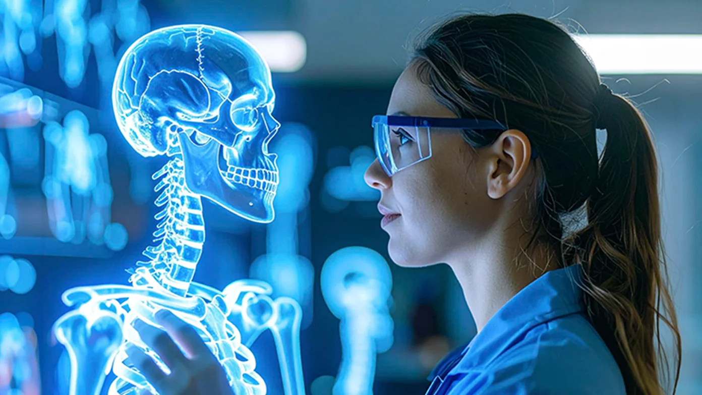
Table of contents
Medical education has always had the same problem: some of the most important things are impossible to see.
You can describe the heart, the brain, or a disease process in a textbook. But understanding how everything fits together is another story.
That's why augmented reality in medical education is becoming part of the curriculum at universities and teaching hospitals worldwide. Instead of imagining anatomy or physiology, students can interact with it.
In this guide, we'll look at where AR fits into medical education, what research says about its effectiveness, and how institutions use custom AR solutions for teaching and clinical training.
Key takeaways
1.
Research shows that immersive learning improves spatial understanding and knowledge retention.
2.
Interactive 3D models help students better understand anatomy, pathology, physiology, and pharmacology.
3.
AR supports every stage of medical education, from classroom learning to surgical training and remote study.
4.
Universities and hospitals can go beyond ready-made apps with custom AR/VR solutions built around their own curriculum.
What is AR in medical education?
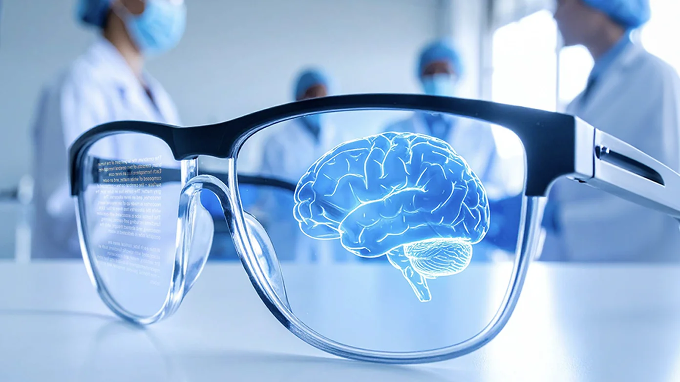
Augmented reality is a technology that overlays digital content like 3D models, animations, and instructions onto real environments. It’s made possible by devices such as AR glasses, smartphones, and tablets. Today, many top medical schools, including Stanford and Columbia University, are already using AR in their teaching toolkit to make lessons more visually engaging for students.
AR in medical education is most often applied through two key scenarios:
-
Displaying hints over mannequins or real bodies
During anatomy lessons, students can wear AR glasses to see labels describing certain systems or organs projected directly onto mannequins or cadavers. This helps to connect what they’ve learned in theory to what they’re seeing and touching in practice.
-
Projecting 3D models and animations in study environments
Using tablets or smartphones with AR-enabled software, students can explore detailed 3D models of organs and body systems in a virtual space. While these models aren’t projected onto real objects like with AR glasses, they offer powerful tools for zooming, rotating, and studying structures from different angles.
Integrating AR in the medical education journey
Learning medicine is not just about memorizing facts. AR supports this journey by bringing more visualization and interaction into classrooms, remote learning, and clinical training.
AR in classroom learning
In classroom settings, AR is turning traditional lectures into immersive learning experiences. Instead of just watching slides, students can use tablets with anatomy apps supporting AR to explore complex topics like organ systems or disease progression in 3D. Even better, with AR glasses, they can see labeled anatomical structures projected right onto mannequins or physical models — muscles, organs, blood vessels, nerves, all layered in front of their eyes. It’s like turning the classroom into a living anatomy lab.
Case Western Reserve University has leveraged this AR-driven approach with the HoloAnatomy app, which teaches anatomy without the need for cadaver labs. Similarly, at Jagiellonian University Medical College in Poland, AR glasses are part of the toolkit in anatomy classes, giving students a more intuitive understanding of spatial relationships in the human body. These tools are especially handy for visual learners and significantly reduce the learning curve for complex medical topics.
AR in remote learning
Augmented reality in healthcare education is also powerful when it comes to remote or hybrid learning environments. With nothing more than a smartphone or tablet, students can project 3D anatomical models right onto their desk at home and manipulate them to explore every angle, layer, and detail.
Many AR platforms also support collaborative learning. For instance, students from different locations can interact with the same virtual model in real time, discussing features, comparing findings, or working through clinical scenarios together. The University of Edinburgh piloted such experiences during the pandemic, allowing medical students to explore anatomy via an AR-based learning platform. It helped students better understand the spatial relationships of pelvic anatomy by projecting interactive 3D models aligned with CT scan data. Available on phones, tablets, or AR headsets, the tool was a great help for both in-person and remote learning scenarios.
AR in advanced surgical training

In live surgical settings, AR technology empowers experienced surgeons to master complex procedures. By overlaying critical information, such as 3D reconstructions of patient anatomy, directly onto their field of view, AR enhances precision and supports more informed decision-making. For instance, Stanford Medicine has integrated AR headsets into the operating room, allowing physicians to visualize complex anatomical structures during procedures. This integration aids in precision and decision-making, ultimately enhancing patient outcomes.
While the primary application of AR in surgery focuses on assisting surgeons, it also offers benefits for medical trainees. By participating in AR-assisted procedures, students gain exposure to advanced surgical techniques and decision-making processes in real-time. This immersive experience provides a deeper understanding of spatial relationships and procedural workflows that are challenging to convey through traditional educational methods.
Subject-specific use cases of AR in the student’s curriculum
Different medical subjects present distinct learning challenges, and the use of AR in medical training helps address them in highly targeted ways. Let’s discuss how it supports learning in specific disciplines.
Anatomy: displaying human organs and tissues
Studying anatomy is one of the most challenging aspects of medical education. It’s dense, highly visual, and often hard to grasp through 2D images. Students need to not only memorize hundreds of structures but also understand how they’re positioned and connected. The interactive nature of AR is a lifesaver here. It helps students explore organs and tissues layer by layer, rotate structures, and view them in context. Thus, students build both spatial awareness and long-term retention.
The interactive nature of AR is a lifesaver here. It helps students explore organs and tissues layer by layer, rotate structures, and view them in context. Thus, students build both spatial awareness and long-term retention. Solutions like VOKA 3D Anatomy and Pathology bring medically accurate 3D models into the classroom, allowing students to examine structures directly on physical surfaces through AR.
Another example of augmented reality in anatomy education is an AR-based vein finder, Hellovein. This device projects real-time visuals over the patient’s skin to show where veins are, which improves accuracy when drawing blood. Tools like this are especially valuable during specialized courses, such as phlebotomy training, helping students build confidence and precision in essential clinical skills.
Pathology: understanding disease development
Pathology studies require visualizing the organ, tissue, and sometimes cellular changes provoked by the disease. With AR in medical education, students can explore how conditions like tumors, atherosclerosis, or organ damage develop over time in a clear visual format. Whether it's a common diagnosis or a rare case study, AR makes it easier to see the progression and connect clinical symptoms to structural changes.
Platforms like VOKA support this approach with detailed 3D pathology models. Students can compare healthy and diseased organs side by side, zoom in to explore damaged areas, and watch how illnesses progress in interactive AR mode. This level of visual clarity gives future doctors a safe space to study complex or uncommon cases they might encounter in real-world clinical practice.
Physiology: visualizing dynamic processes & procedures

Physiology is all about movement: blood pumping, lungs expanding, neurons firing. And let’s face it, those dynamic processes are tricky to understand through still images. That’s where AR really shines, as it gets students “inside” the process. They can watch 3D animations in augmented reality that bring organs and systems to life, seeing how everything works together in real time. Instead of imagining how the body works, AR shows it right in front of their eyes.
But AR doesn’t stop at just showing how the human body works. It also lets students practice. A great example is the AR defibrillator, which places students in a realistic cardiac arrest scenario where they practice hand placement, deliver a simulated shock, and watch how the heart’s rhythm responds to each step. It’s a hands-on way to build confidence when interacting with physiological processes in high-pressure situations.
Pharmacology: mechanism of action made visible
In pharmacology, it’s crucial for students to understand how drugs travel through the body, interact with cells, and produce specific effects at the microscopic level. For that purpose, students can leverage VOKA’s dedicated microanatomy section to dive into the world of cells with AR. When studying topics at the macroscopic level, being able to zoom in and explore more detailed 3D structures (like tissues or individual components) helps deepen understanding, especially in areas like pharmacology where precision matters.
Students can also take it a step further by watching the mechanism of action (MoA) animations through AR glasses. These immersive visuals show exactly how molecules bind to receptors, how ion channels open, or how enzymes are inhibited. Apart from being a powerful visualization aid, such 3D medical animations also help future doctors communicate drug mechanisms more clearly to patients and colleagues.
Benefits of AR for medical students

AR is not just about making medical education look more impressive. It solves some very practical problems students face every day: understanding complex structures, remembering what they learn, and feeling more confident when applying knowledge.
Research also supports these benefits. Let’s look at where AR makes the biggest difference.
Interactive exploration with better visibility
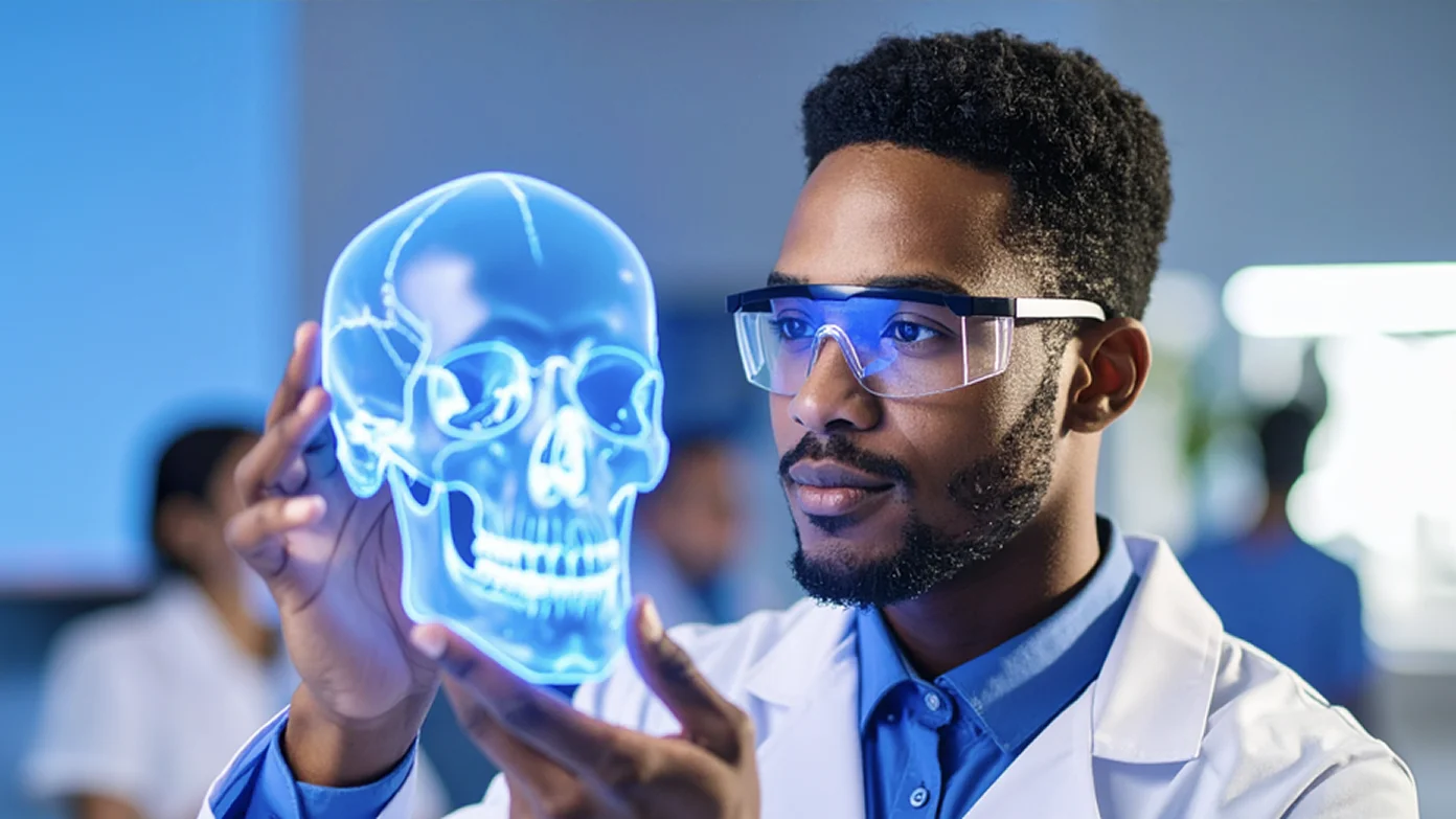
Looking at a 2D image and understanding a 3D structure are two completely different things.
AR helps students explore anatomy and pathology from every angle. They can rotate models, examine different layers, and see how structures connect in real space, instead of trying to build the picture in their heads.
A study published in Anatomical Sciences Education found that medical students who learned anatomy with mobile AR achieved higher learning outcomes while experiencing lower cognitive load compared to traditional approaches.
Improved knowledge retention
People remember things better when they actively interact with what they are learning.
Instead of memorizing structures from static images, AR allows students to explore, manipulate, and revisit complex concepts in a more hands-on way. This active learning approach helps turn short-term memorization into deeper understanding.
Research on AR in medical education shows that immersive and interactive tools can improve engagement and support better learning outcomes.
Boosted confidence in clinical decision-making
By providing real-time visual cues and contextual information, AR helps students make more informed choices during practical exercises.
Seeing relevant anatomical data, step-by-step guidance, or diagnostic hints right in their field of view reinforces correct reasoning and reduces hesitation.
Over time, this repeated support strengthens students’ clinical judgment and builds confidence in applying knowledge under pressure.
Custom AR/VR development: why choose VOKA for your institution
Off-the-shelf AR apps can be a great starting point. They give students access to 3D models and help make lessons more interactive.
But universities, hospitals, and training centers often need something more specific. Maybe you want to teach a rare pathology that is not available in standard libraries. Or you need a surgical simulator built around your training program. Or you look for AR modules that fit directly into your existing LMS.
That’s where custom AR/VR development makes a difference.
With a custom solution, institutions can create immersive learning environments designed around their own curriculum, clinical workflows, and educational goals.
VOKA combines medical expertise with 3D development capabilities to build custom AR/VR experiences, from anatomy and pathology visualizations to advanced training tools.
If your institution has a specific training challenge, contact VOKA so we can help turn it into an interactive solution.
FAQ
1. What is augmented reality in medical education?
Augmented reality in medical education is the use of AR technology to overlay digital 3D models, animations, and labels onto real-world environments. With AR glasses, tablets, or smartphones, medical students can explore anatomy, physiology, pathology, and even pharmacology in an interactive and immersive way.
2. How does augmented reality improve medical training?
Augmented reality for medical training enhances student learning by making anatomy and physiology more interactive, allowing learners to visualize structures in 3D, and practice clinical skills in safe, simulated environments.
3. What are the benefits of using AR in healthcare education?
The benefits include better visualization of complex structures, improved knowledge retention, enhanced confidence in clinical decision-making, and more engaging training sessions.
4. Is augmented reality the future of medical education?
Yes, AR in medical education is rapidly becoming a core learning tool. Universities and hospitals are adopting AR for anatomy classes, remote learning, and surgical training. As the technology develops, it’s expected to play an even bigger role in preparing future healthcare professionals.
Make VOKA your preferred source
See more VOKA articles in Google Search

Share
Table of contents

Thank you for your comment!
Your comment has been submitted for moderation and will be published soon. We'll email you once it’s live.